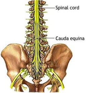
 The words, cauda equina, used to describe the bottom part of the spinal cord literally mean “horse’s tail.” Those Latin and Greek fathers of western medicine called it like they saw it.
The thin nerves emerge from the bottom of the spinal cord proper and are bundled together like hair in the tail of a horse. Every human has a cauda equina running inside the bones of their spine. It begins near the bottom of the ribs.
A tumor in this region of the spine can be tricky to remove. It requires a surgeon with good technique and a delicate touch.
Dr. Paul McCormick, director of The Spine Hospital at The Neurological Institute of New York, is a specialist in removing this kind of tumor and he shared his technique in a surgical video published recently by the Journal of Neurological Surgery: Neurosurgical Focus.
The patient in Dr. McCormick’s video is a man in his early thirties who has been having back and leg pain for a couple of months. His pain turns out to be due to a nerve tumor, or schwannoma, growing in his cauda equina.
The tumor in this patient’s cauda equina means bad news for one of those nerves.
The nerve has two strands: a sensory strand for carrying sensory information from the body to the brain; and a motor strand for carrying instructions about movement from the brain to the body. The sensory strand of this nerve has the tumor. It cannot be saved.
But with great care, the movement strand of the patient’s nerve can be saved. The operation is delicate, though. Both nerve strands are tucked together inside one membrane. Swelling from the tumor means that the movement nerve has grown stuck to the tumor. Separating and removing the entire tumor without damaging the motor nerve requires surgical expertise and a thorough knowledge of the area’s anatomy.
The video is intended to teach other spinal surgeons, so Dr. McCormick’s skill as both a surgeon and a teacher is on display. He uses video from his surgical microscope as well as diagrams that explain how the nerves, membranes, and blood vessels are arranged. He lets the viewers know about the most common challenges seen in this type of surgery, and how to overcome them. And he describes the nerve testing he conducts throughout the operation.
In the end, the operation is a success. Dr. McCormick’s patient wakes up tumor-free, and with all his motor nerves intact.
The video is available on YouTube. But be aware: it is a graphic look at spinal surgery through the lens of an operating microscope. Interested adults can watch the video here: Microsurgical Resection of Cauda Equina Schwannoma with Nerve Root Preservation.
The video is one of several spine surgery videos that Dr. McCormick produced for Journal of Neurological Surgery. Read about Dr. McCormick’s other patients and videos below. Read about his role editing the overall project here.
Find out more about Dr. McCormick on his bio page here.
Honors and Awards 2026 (Pictures)
Dr. McCormick named as one of Castle Connolly’s Top United States and New York Doctors/Neurosurgery for 25 consecutive years.