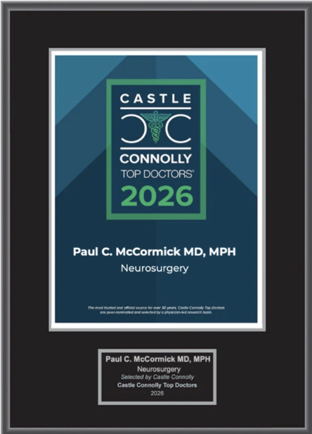
Dr. McCormick named as one of Castle Connolly’s Top United States and New York Doctors/Neurosurgery for 25 consecutive years.
| Header | Text |
| Overview | In microsurgery, a surgeon uses an operating microscope and specialized instruments to perform surgery on small or delicate structures. Operating microscopes are designed to provide bright light and greatly magnify the surgical area. This allows the trained neurosurgeon to be more precise and effective in operating the delicate structures around and in the spinal cord and nerve roots. Many operating microscopes also display a high-resolution image on a screen in the operating room, and some allow recording of the procedure. See below for some videos of spinal microsurgery conducted by Spine Hospital Director Dr. Paul C. McCormick who produced the videos as teaching aids for other neurosurgeons. They are available for public viewing, but keep in mind that they contain close-up footage of spine surgery.
Microsurgery may be used to remove a tumor or tangle of blood vessels in the spinal cord, a procedure called microsurgical spinal cord tumor resection. It may be used to remove part of a damaged disc, as in a cervical microdiscectomy or a lumbar microdiscectomy. In fact, it may be used as part of many surgeries, including minimally invasive surgery.
|
| Preparing for Your Appointment | The world-class neurosurgeons at The Spine Hospital at the Neurological Institute of New York continue a long tradition of skill, care, and expertise that result in the best possible surgical outcomes for our patients. Drs. Paul C. McCormick, Michael G. Kaiser, Peter D. Angevine, Alfred T. Ogden, Christopher E. Mandigo, Patrick C. Reid and Richard C.E. Anderson (Pediatric) are experts in microsurgical spine treatments. |
Helpful Surgery Overviews
Dr. McCormick will choose the treatment method specific to each patient and situation. Some of the condition’s treatment options may be listed below.
Recent News
Dr. McCormick named as one of Castle Connolly’s Top United States and New York Doctors/Neurosurgery for 25 consecutive years.

Dr. McCormick participated as both a member of the expert panel as well as a co-author of recently published randomized study on the treatment of grade 1 degenerative spondylolisthesis. This condition commonly occurs in adult patients and is subject to numerous types...
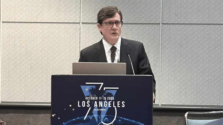
Dr. McCormick was invited to present his experience on the evaluation and microsurgical management of intramedullary spinal cord tumors. Dr. McCormick described his techniques and illustrated on video the safe and complete microsurgical resection of both an...