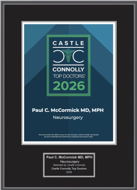
Dr. McCormick named as one of Castle Connolly’s Top United States and New York Doctors/Neurosurgery for 25 consecutive years.
| Header | Text |
| What is Spinal Cord Tumor Resection? | Spinal cord = the bundle of nerves that connect the brain to the rest of the body Tumors may evolve on any nerve in the body, but tumors on the spinal cord and its associated structures are rare. When they do occur, the majority of spinal cord tumors are not cancerous and don’t often spread beyond the spinal canal. Therapies such as radiation or chemotherapy often have little effect on these tumors. Thus surgical resection (removal of the tumor) may often be the preferred treatment for a spinal cord tumor. Spinal cord tumor resection is performed for both adult and pediatric patients who have tumors on the spinal cord. At The Spine Hospital at the Neurological Institute of New York, we specialize in spinal cord tumor resection. |
| When is this Procedure Performed? | Most spinal tumors are not cancerous and don’t spread. Very small spinal tumors may not even cause symptoms, and therefore may not need to be removed. But when these tumors compress the spinal cord or spinal nerves, a patient may experience weakness, numbness, loss of balance, difficulty walking or talking, or loss of bowel or bladder control. In these cases, microsurgical removal may be the best treatment option. Together, neurosurgeon and patient (or patient’s guardian) will be able to assess whether surgery is the preferred treatment. The most common tumors developing from within the spinal cord are ependymomas, astrocytomas or hemangioblastomas. Tumors such as schwannomas, neurofibromas, or meningiomas may also develop from supporting tissues outside the spinal cord. |
| How is this Procedure Performed? | The procedure is performed under general anesthesia, which means the patient is unconscious during the procedure. Before and during the operation, the surgeon and neurophysiologists conduct electrophysiological monitoring of spinal cord function. This monitoring includes SSEPs (somatosensory evoked potentials) and MEPs (motor evoked potentials), tests that show the electrical impulse of a signal passing through the spinal cord and spinal nerves. First, the neurosurgeon makes an incision in the skin overlying the tumor. The soft tissues and muscles are moved aside to expose the back of the spine, and the laminae (superficial spinal bones) are removed to access the spinal canal. In rare circumstances, when the tumor is on the front of the spinal cord, the neurosurgeon may access the spinal canal from the front or the side of the spine. Within the spinal canal is a tissue-lined compartment that contains the spinal cord and nerves, which are bathed in cerebrospinal fluid. The tissue lining of this compartment is known as the dura. The dura is opened to expose the spinal cord and nerves. Using the high magnification and illumination of the operating microscope, our neurosurgeons apply microsurgical techniques to carefully dissect the tumor from the surrounding structures to the greatest extent possible for safe removal. Electrophysiological monitoring continues during this period to continue to oversee safety of the spinal cord. Once the tumor is removed the dura is sutured or stitched closed. A water-tight closure of the dura is important in order to avoid leakage of the cerebrospinal fluid. Spinal fusion is rarely needed following removal of these tumors. The soft tissues overlying the spine are then closed in multiple layers and the skin closed with either staples or a nylon suture. The incision is then covered with a gauze bandage. |
| How Should I Prepare for this Procedure? | Make sure you understand the goals of the procedure as well as what you or your child can expect after surgery. Some people find it helpful to write down their questions and bring the list of questions to their appointments. Make sure to tell your doctor about any medications or supplements that you or the patient are taking, especially medications that can thin the blood such as aspirin. Your doctor may recommend you or the patient stop taking these medications before the procedure. To make it easier, write all the medications down before the day of surgery. Be sure to tell your doctor if you or the patient have an allergy to any medications, food, or latex (some surgical gloves are made of latex). On the day of surgery, remove any nail polish or acrylic nails, do not wear makeup and remove all jewelry. If staying overnight, bring items that may be needed, such as a toothbrush, toothpaste, and dentures. You will be given an ID bracelet. It will include your or the patient’s name, birthdate, and surgeon’s name. |
| What Can I Expect After the Procedure? | How long will I stay in the hospital? Will I need to take any special medications? Will I need to wear a collar or a brace? When can I resume exercise? Will I need rehabilitation or physical therapy? Will I have any long-term limitations due to microsurgical tumor removal? |
| Preparing for Your Appointment | Drs. Paul C. McCormick, Michael G. Kaiser, Alfred T. Ogden, Christopher E. Mandigo, Patrick C. Reid, Richard C. E. Anderson (Pediatric), and Neil A. Feldstein (Pediatric) are experts in spinal cord tumor resection.
|
Helpful Surgery Overviews
Dr. McCormick will choose the treatment method specific to each patient and situation. Some of the condition’s treatment options may be listed below.
Recent News
Dr. McCormick named as one of Castle Connolly’s Top United States and New York Doctors/Neurosurgery for 25 consecutive years.

Dr. McCormick participated as both a member of the expert panel as well as a co-author of recently published randomized study on the treatment of grade 1 degenerative spondylolisthesis. This condition commonly occurs in adult patients and is subject to numerous types...
Dr. McCormick was invited to present his experience on the evaluation and microsurgical management of intramedullary spinal cord tumors. Dr. McCormick described his techniques and illustrated on video the safe and complete microsurgical resection of both an...