| Header | Text |
| Summary | Spinal = having to do with the spine
Stenosis = narrowing
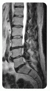
 MRI of Lumbar Spinal Stenosis Multiple level disc bulging and spinal canal narrowing has obstructed the flow of the clear white spinal fluid within the lower canal.
Spinal stenosis is a narrowing of the spinal canal, the bony structure that encloses the spinal cord and the nerve roots.
Spinal stenosis usually develops gradually due to age-related spinal degeneration. It is most common in people over 50 years of age, and is widely recognized as a significant source of disability.
Most cases of spinal stenosis occur in the lumbar spine (lower back). However, stenosis may also occur in the cervical spine (neck). Rarely, spinal stenosis occurs in the thoracic spine (mid- and upper back).
|
| Symptoms | Symptoms of spinal stenosis are produced when the narrowing spinal canal compresses the spinal cord or nerve roots. Specific symptoms depend on the location and severity of the stenosis.
Cervical spinal stenosis may cause the following symptoms:
- numbness or clumsiness of hands
- balance and coordination problems, such as leg stiffness or tripping while walking
- weakness or incoordination of the arms and hands
- difficulty with fine motor control of hands such as buttoning, tying, writing and typing
- difficulty with bladder or bowel control
Lumbar spinal stenosis may cause the following symptoms:
- low back pain
- pain, tingling or weakness/heaviness in one or both legs that becomes worse with standing or walking. These symptoms are often relieved with sitting or leaning forward, as while pushing a shopping cart. This is called neurogenic claudication, and it occurs in 90% of spinal stenosis cases.
- weakness, numbness or pain that radiates to the buttocks and legs. These are symptoms of radiculopathy, or compression of a lumbar nerve root.
|
| Causes and Risk Factors | As many as 90% of reported cases of spinal stenosis result from degenerative changes that occur with aging. This degeneration, called spondylosis, can lead to bone spurs that may narrow the spinal canal. Other possible contributors to spinal stenosis include the thickening of ligaments within the spinal canal; the degeneration, herniation or bulging of intervertebral discs; and the formation of synovial cysts (fluid-filled sacs in the joints).
Although other problems such as fractures, tumors, infection, systemic bone diseases, or conditions present at birth can cause narrowing of the spinal canal, the term “spinal stenosis” is generally reserved for degenerative causes.
|
| Tests and Diagnosis | The diagnosis of spinal stenosis begins with a complete history and physical examination. The doctor will determine what symptoms exist, what makes them better or worse, and how long they have been present. A neurological examination that demonstrates abnormalities in the strength, sensation and reflexes of particular parts of the body may provide objective evidence of spinal cord or nerve root compression caused by spinal stenosis.
In addition to taking a history and conducting a physical examination, the doctor may order the following exams to help diagnose spinal stenosis:
- X-ray (also known as plain films) –test that uses invisible electromagnetic energy beams (X-rays) to produce images of bones. Soft tissue structures such as the spinal cord, spinal nerves, the disc and ligaments are usually not seen on X-rays, nor on most tumors, vascular malformations, or cysts. X-rays provide an overall assessment of the bone anatomy as well as the curvature and alignment of the vertebral column. Spinal dislocation or slippage (also known as spondylolisthesis), kyphosis, scoliosis, as well as local and overall spine balance can be assessed with X-rays. Specific bony abnormalities such as bone spurs, disc space narrowing, vertebral body fracture, collapse or erosion can also be identified on plain film X-rays. Dynamic, or flexion/extension X-rays (X-rays that show the spine in motion) may be obtained to see if there is any abnormal or excessive movement or instability in the spine at the affected levels.
- Magnetic resonance (MR) imaging – provides detailed images of soft tissues like the spinal cord and nerve roots. As a result, MRIs are very helpful in determining the location and severity of the stenosis and in identifying spinal cord or nerve root compression.
- Computed tomography scan (CT scan) – this scan uses X-rays and a computer to provide images that are more detailed than general X-rays.
- Myelogram – X-rays and CT scans taken after a dye is injected into the spinal canal. This test is useful in patients who have had previous surgery or who have another condition, such as scoliosis.
|
| Treatments | Before surgery is considered, a doctor will typically recommend nonoperative treatments. These measures may include anti-inflammatory medications, physical therapy, weight control, or pain management techniques such as epidural spinal injections. A doctor or physical therapist may provide an exercise program or instruction on proper posture.
When nonoperative treatments don’t help or stop working, surgery often provides relief. For patients with persistent neurogenic claudication, a decompressive lumbar laminectomy may be recommended. In a laminectomy, a surgeon removes a section of vertebral bone called the lamina. This makes more room for the spinal cord and nerve roots in the spinal canal. Other surgeries may be recommended to remove portions of the joints, disc, or ligaments that compress the spine or nerve roots.
The surgeon assesses the likely effect of any such surgery on the stability of the spine. If the spine is or may become unstable, a stabilization and fusion procedure may be considered.
|
| Preparing for Your Appointment | Drs. Paul C. McCormick, Michael G. Kaiser, Peter D. Angevine, Alfred T. Ogden, Christopher E. Mandigo, Patrick C. Reid and Richard C.E. Anderson (Pediatric) are experts in treating spinal stenosis. They can also offer you a second opinion.
|


