Back in 1910, Dr. Charles Elsberg performed the first surgery at the Neurological Institute of New York. That operation was the removal of an “intramedullary” spinal cord tumor, and it was truly a groundbreaking procedure.
An “intramedullary” spinal cord tumor grows “intra” (within) the “medulla” (middle) of the spine. Such tumors grow in among the spinal cord’s delicate nerve fibers. These fibers and the tumor are all bundled together, squeezed inside the spinal cord’s protective membrane.
Back in 1910, almost nobody operated to remove these tumors. Moving the nerve fibers aside and resecting the tumors usually caused too much damage.
But at the Neurological Institute, Dr. Elsberg pioneered the first safe and reliable way to remove such tumors. His method was to perform two surgeries, on two separate days. In the first surgery, he operated down to the spinal cord, and made an incision above the tumor in the spinal cord’s protective membrane. The protective membrane surrounds and contains the spinal cord. Then he closed the skin incision, leaving the deep incision over the tumor open.
A week later, he operated again. The pressure of the tumor would force much of the tumor out through the membrane incision. This made it more feasible for the doctor to remove the tumor. (For more on Dr. Elsberg and his interesting method, see our blog post here.)
More than 100 years later, a whole lot has changed in neurosurgery. Today, the Neurological Institute is home to our Department of Neurosurgery. Surgeons routinely remove intramedullary spinal cord tumors. But one thing is still the same: doctors here are still at the forefront when it comes to spinal cord tumor surgery.
 The Spine Hospital at the Neurological Institute of New York’s Director Dr. Paul McCormick is one of today’s leading experts in removing intramedullary spinal cord tumors. He doesn’t use Dr. Elsberg’s two-stage method, though. He performs these delicate, exacting surgeries in one stage, using instruments that Dr. Elsberg could only have imagined: microsurgical implements, lasers, and ultrasonic aspirators. He can tailor his instruments and approach to remove spinal tumors of many different types.
Not only have the last hundred years brought a host of new surgical methods, but they have also brought new instructional methods. Dr. McCormick recently edited a spinal tumor video series for the Journal of Neurosurgery. The series, intended as an educational tool for other neurosurgeons, is freely available to everyone on Youtube.
Each video presents a case study with diagnostic images, a voice-over of the procedure, and plenty of footage from Dr. McCormick’s surgical microscope. You can read about one of his intramedullary spinal cord tumor videos in our post here, or watch the video here.
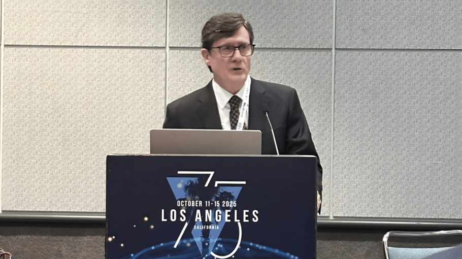
Dr. McCormick also helps other neurosurgeons learn about the latest developments in neurosurgery. At this year’s meeting of the American Association of Neurological Surgeons, Dr. McCormick spoke on a panel about intramedullary spinal cord tumors.
The seminar lasted two hours, and it covered how to assess technical advances in surgery, how to reduce operative complications, and how to identify the best operative techniques for each type of tumor.
The last hundred years have seen incredible advances in intramedullary spinal cord tumor surgery. It’s exciting to think what changes the next hundred years will bring to The Spine Hospital at the Neurological Institute of New York.
Learn more about Dr. McCormick on his bio page here.
*Top image courtesy of the Archives & Special Collections, Columbia University Health Sciences Library