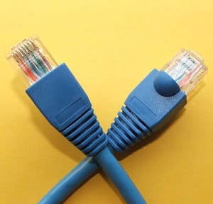
 Imagine the cable that brings Internet access to your home. Inside that cable are many small wires. Outer layers of the cable surround and protect those individual wires, bundling them all together.
Now imagine the human spinal cord. It too is composed of tiny “wires,” or delicate nerve fibers, bundled together inside a series of protective membranes.
Outside the spinal cord, the bony cage of the spinal column provides even more protection. Vertebra are stacked on top of each other like marshmallows on a stick, a hole down the center of each providing safe passage for the spinal cord.
But sometimes a threat comes from inside the spinal cord–not outside. Then a surgeon must open all those layers of protection and access the delicate nerves of the spinal cord. This process is called intramedullary spinal surgery, and it’s incredibly exacting.
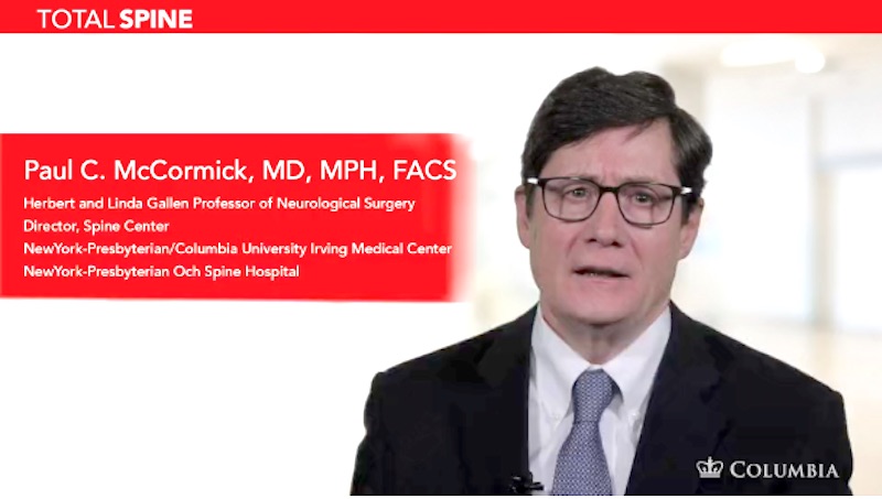
Dr. Paul McCormick is a leading expert on intramedullary spinal surgery. In a series of videos, he aims to share his knowledge and experience with other neurosurgeons.
In this video, his patient is a woman in her 50s who suddenly developed arm pain and weakness. She was diagnosed with a cavernous malformation, a tangle of irregularly-shaped blood vessels that are prone to dangerous leaking. Her cavernous malformation is high up in her neck, just below the base of her skull. It is in the intramedullary space, right in among those delicate nerve fibers. And it must be removed.
The first attempt at removing this cavernous malformation had to be called off. During that operation, vertebral bone was removed and the spinal cord was closely monitored using techniques called Somatosensory Evoked Potentials (SSEP) and Motor Evoked Potentials (MEP). These techniques measure electrical activity in the spinal cord. Careful SSEP and MEP monitoring showed that the spinal cord was not responding safely to the next part of the surgery. To avoid injuring the spinal cord, the surgery was called off before the cavernous malformation could be removed.
In this video, Dr. McCormick works with his colleague Dr. Jeffrey Bruce, Director of Skull Base Surgery. The surgeons approach the problem from, literally, a different angle. They use a “far lateral” approach, coming at the problematic area from the side. With this video, they will help other neurosurgeons who need to use this non-traditional approach to the area.
In the video, Dr. McCormick shows where vertebral bone was removed in the patient’s first surgery. Though the surgeons now approach from a different angle, they can still use this area to access the spinal cord.
Once the spinal cord is open, Dr. McCormick works in the intramedullary space with meticulous care, monitoring the spinal cord constantly. Piece by piece, he extracts the cavernous malformation. Finally, he inspects the spinal cord with a microscope under high magnification: none of the cavernous malformation remains.
Then it’s time to put the spinal cord’s protective membranes back in place. Dr. McCormick uses a microscope and micro-instruments to stitch closed the spinal cord’s outer membrane. Before it was opened by his surgical incision, this membrane sealed in the cerebrospinal fluid that bathes the spinal cord. When he’s through stitching, the membrane is watertight once again.
After this angle of approach to the spinal cord, patients can sometimes experience shoulder weakness. This is because the nerves that go to the shoulder and arm pass through this area. This patient experiences shoulder weakness, which improves over the course of the next four months. The patient’s original weakness improves, too.
Removing a cavernous malformation is a delicate business. But with experience, meticulous care, and unflagging attention to detail, surgeons can do it as safely as possible. And with this video, they can benefit from Dr. McCormick’s and Dr. Bruce’s experience.
Dr. McCormick’s video on cavernous malformation surgery is available for the public to watch. Dr. McCormick notes that patients are often interested in seeing a video of “their” type of surgery. But be aware: this video is an educational resource for neurosurgeons. It contains detailed surgical footage, and should only be watched by interested adults. Watch the video here.
Learn more about Dr. McCormick on his bio page here.
Image credit: “Pkuczynski RJ-45 patchcord”. Licensed under CC BY-SA 3.0 via Wikimedia Commons


