
The patient is a 63-year-old woman. Her trouble with walking has been getting worse… and worse… and worse.
The trouble turns out to come from a tumor in her spine. It’s a schwannoma, a nerve tumor that is benign. (That is, it’s not going to spread.)
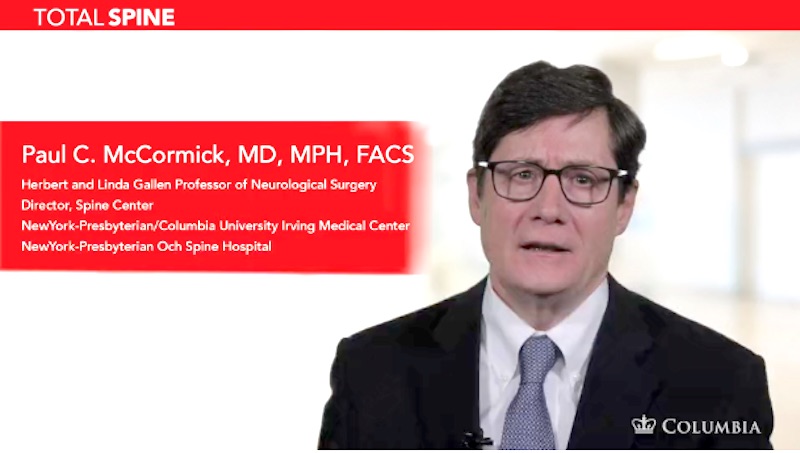
But this tumor can’t stay where it is. It is compressing the delicate nerves in her spinal cord, leading to the woman’s gait problems. Left unchecked, the tumor would cause progressively more problems. But The Spine Hospital at the Neurological Institute Director Dr. Paul McCormick will remove the tumor, sparing the nerves of the spine.
Dr. McCormick is a world-renowned spinal surgeon. To share his expertise with other surgeons, Dr. McCormick produced an educational video about this patient’s diagnosis, surgery, and recovery.
In the video, Dr. McCormick shows the patient’s MRI. The tumor is easy to see. It is about the size and shape of two gumdrops. It is so large that it severely compresses the spinal cord, shoving it out of the way and—Dr. McCormick points out—rotating that important bundle of nerves to the left.
As Dr. McCormick explains, this rotation leaves a useful narrow space on the right. With the spinal cord safely out of the way, he can approach through that narrow corridor to get at the tumor.
And this he does. He shows footage recorded through his operative microscope. Working on the first “gumdrop,” he narrates his techniques and gives tips for safe tumor removal.
While breaking up and removing small parts from inside the tumor, he notes, “Care must be taken to limit further manipulation of an already severely compressed spinal cord during this part of the [removal].” In other words, the famed “surgeon’s hands” are especially necessary here. It is imperative not to jostle or compress the spinal cord…even though the tumor he’s excavating is shoved right up alongside it.
Finally, he has removed enough bulk from inside the tumor. Now he can compress it and slide it out of the narrow corridor. Beneath the tumor is its final connection to the patient’s body–the thin nerve that the large tumor grew from. Dr. McCormick snips this connection, and the first part of the schwannoma removal is complete. Then he proceeds to the next tumor nodule and removes it as well. Finally, he explains how he closes up the incisions.
The patient had a great recovery. She was up and around two days after surgery, and went home from the hospital two days after that. Her gait improved significantly. And one year later, there is no evidence of any tumor.
This patient is grateful for her surgeon’s skill and expertise. His steady hands got her back on her feet.
You can watch the video here. But be aware, this video contains a detailed, close-up look at spine surgery. It should only be watched by interested adults.
Read more about Dr. McCormick on his bio page here.
Watch more Spinal Surgery Videos by Dr. Paul McCormick here.
Image credit: [Werneuchen] / Wikimedia Commons
Posted on Oct 9, 2015 by Department Author
