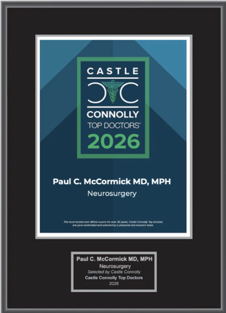
Dr. McCormick named as one of Castle Connolly’s Top United States and New York Doctors/Neurosurgery for 25 consecutive years.
| Header | Text |
| Summary | Cavernous = Refers to the sacs of pooled blood inside the malformation A cavernous malformation is an abnormal collection of tiny blood vessels called capillaries. Cavernous malformations can occur anywhere in the body, and in many locations they are harmless. In the spinal cord, however, they can present a problem. At the Spine Hospital at the Neurological Institute of New York, we specialize in cavernous malformations of the spinal cord. The capillaries in a cavernous malformation have especially thin walls. They are prone to leaking and bulging outward repeatedly, forming sacs where blood pools and clots. This collection of sacs gives cavernous malformations a characteristic “mulberry-like” appearance. |
| Symptoms | Most spinal cavernous malformations cause no symptoms at all. Asymptomatic (symptomless) spinal cavernous malformations may not need treatment. Like other spinal lesions, cavernous malformations may cause symptoms when they compress and damage the delicate nerves of the spinal cord. The mass of the malformation itself can cause such compression, as can the swelling caused by a hemorrhage of blood from the abnormal vessels. Exact symptoms depend on the size and location of the cavernous malformation. Symptoms may include pain, weakness, numbness, tingling or clumsiness in the extremities. |
| Causes and Risk Factors | Spinal cavernous malformations are rare, and their exact causes are still being researched. In some cases, cavernous malformations run in families. People with the familial (genetic) form of cavernous malformation are likely to have more than one malformation, and likely to have malformations that are candidates for surgical treatment. The relevant mutation is inherited in an autosomal dominant pattern–that is, a parent with the disease has a 50% chance of passing it along to each of his or her children. In most other cases, known as sporadic cases, the cause is not genetic. Sporadic cases account for more than 80% of cavernous malformations. They are more likely to be isolated (only one malformation per individual), are not passed on to children, and are less likely to cause symptoms. |
| Tests and Diagnosis | Magnetic resonance (MR) scans are the most useful diagnostic study for cavernous malformations. These scans use large magnets, radiofrequencies, and a computer to produce detailed images of organs and structures inside the body. Computed tomography scans (CT scans, or CAT scans) may also be used. These scans use X-rays and a computer to produce detailed images of bones, muscles, fat and organs. Angiography is rarely used. Angiograms are designed to study blood flow, but since blood flows very slowly through a cavernous malformation, angiograms cannot provide much useful information. Cavernous malformations are known therefore as “angiographically occult” lesions–lesions that are invisible to angiography. Molecular genetic testing is available to confirm the diagnosis of familial cavernous malformation. It is not a useful test in most cases of sporadic cavernous malformation–in those cases, the genetic form is already ruled out by the isolated lesion and lack of family history of the disease. |
| Treatments | The decision to treat a cavernous malformation depends on whether it causes symptoms, the patient’s age and health status, and the risks of surgery. Sometimes a cavernous malformation may simply be observed over time with regular MRI scans. In cases of large or symptomatic cavernous malformations, or malformations with recurrent hemorrhages, surgery to remove the malformation may be considered. Microsurgery, or surgery using an operating microscope and very fine instruments, is the procedure of choice for cavernous malformation removal. Because cavernous malformations do not infiltrate healthy tissue, they usually can be fully removed with microsurgical techniques. Watch Dr. Paul C. McCormick perform this microsurgery by clicking on the video link below. However, spinal cavernous malformations are complex lesions near highly sensitive structures. They should be addressed at a major medical center like The Spine Hospital at The Neurological Institute by experts like our neurosurgeons who are experienced in their treatment. |
| Preparing for Your Appointment | Drs. Paul C. McCormick, Michael G. Kaiser, Alfred T. Ogden, Christopher E. Mandigo, Patrick C. Reid, Richard C.E. Anderson (Pediatric), and Neil A. Feldstein (Pediatric) are experts in treating cavernous malformation. They can also offer you a second opinion. |
Helpful Surgery Overviews
Dr. McCormick will choose the treatment method specific to each patient and situation. Some of the condition’s treatment options may be listed below.
Recent News
Dr. McCormick named as one of Castle Connolly’s Top United States and New York Doctors/Neurosurgery for 25 consecutive years.

Dr. McCormick participated as both a member of the expert panel as well as a co-author of recently published randomized study on the treatment of grade 1 degenerative spondylolisthesis. This condition commonly occurs in adult patients and is subject to numerous types...
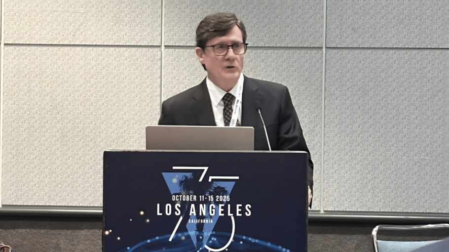
Dr. McCormick was invited to present his experience on the evaluation and microsurgical management of intramedullary spinal cord tumors. Dr. McCormick described his techniques and illustrated on video the safe and complete microsurgical resection of both an...