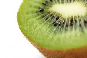
 There are a lot of ways to go after a kiwi. Some people consume the whole thing, skin and all. Others work hard to get out all the green insides without breaking through the thin skin.
If you use this second method, you’ve probably noticed that it’s gotten easier with practice. Well, in kiwis as in spine surgery, experience means a lot.
Dr. Paul McCormick has had years of experience in spine surgery, and he is an expert in this difficult field. In one recent operation, he needed to remove most of an intramedullary lipoma without breaking through its “skin.” The process was reminiscent of kiwi-extraction… except far more difficult, with much higher stakes.
A lipoma is a benign growth of fatty tissue. An intramedullary lipoma is one of these growths inside the spinal cord’s neural tissue, also known as the intramedullary space. In a new video, Dr. Paul McCormick explains the process of removing an intramedullary lipoma from the spinal cord of a 26-year-old man.
Lipomas are not cancer. They will not spread, and they can usually be left in place. But this patient’s lipoma is taking up too much important room, squeezing the delicate spinal nerves. The pressure on the nerves is causing pain in the patient’s neck and a “pins and needles” feeling in his arm. Removing the lipoma will give the spinal nerves the comfortable space they need again.
Dr. McCormick’s goal in this surgery is not to remove the entire growth. Instead, he will leave a small amount behind–a hollowed-out remnant kind of like the rind of a kiwi.
 Why leave some behind? This intramedullary lipoma has actually grown attached to the nerves of the spinal cord. Spinal nerves are delicate, and they do not repair themselves if damaged. A surgeon must balance the need to free up space for the spinal nerves and the need to leave those nerves undisturbed. Dr. McCormick is an expert in surgery involving the spinal cord, so he is highly proficient at finding this balance.
He begins by working on the safe, central portion of the intramedullary lipoma—the part that isn’t touching the spinal cord. Using his surgical microscope and micro-instruments, he removes piece after tiny piece.
Soon, Dr. McCormick explains in the video, a surgeon can bring in a new tool: intraoperative ultrasound. This type of ultrasound can help guide a surgeon as he works ever closer to the spinal nerves.
Finally, Dr. McCormick is satisfied with the removal. He closes up the incisions.
The patient does very well after surgery. He is up and about two days later. His neck pain and “pins and needles” disappear. And an MRI taken 6 months post-surgery shows that only a thin layer of lipoma remains. The removal was a success.
Dr. McCormick produced this video as a teaching aid for other neurosurgeons. Its thorough technical details will help neurosurgeons who have less experience with intramedullary lipoma surgery. But Dr. McCormick had another reason for making this video, too. He believes that some patients might want to watch the video. He says he is surprised by how many people ask to watch a video of “their” type of surgery.
The video is available to watch here. But be aware: this is a technical video that contains a lot of close-up footage of spine surgery. It is intended primarily as a teaching tool for other neurosurgeons. It should be watched only by interested adults.
Read about (or watch) Dr. McCormick’s other fascinating videos, collected on his videos page here.
Learn more about Dr. McCormick on his bio page here.
Image credit: “Kiwi Fruit” [Jeremy Knight] / Flickr
Kiwi eaters: many people find that using a spoon is the easiest way to peel a kiwi. Video demonstration–suitable for all ages–here.