 Step into an operating room at The Spine Hospital at the Neurological Institute of New York today and you’ll find all kinds of high-tech equipment: Digital microscopes that illuminate and magnify the surgical field. Lasers that precisely cut away abnormal tissue. Ultrasonic aspirators that break up and remove tissue fragments.
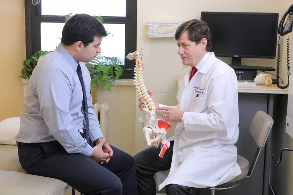
Spine Hospital Director Dr. Paul McCormick uses this equipment and more to perform an extremely delicate kind of spine surgery: removing tumors from inside the spinal cord itself.
Tumors inside the spinal cord are called intramedullary tumors. Their location within the spinal cord means that in order to reach them, Dr. McCormick must move aside the long fibers of the spinal cord. Since injuries to the spinal cord do not always heal, Dr. McCormick and other surgeons who work with the spinal cord must be exceedingly skilled and careful.
They use electrophysiological equipment and computers to monitor the health and safety of the spinal cord while they operate. Together, the doctors’ surgical skill and advanced technology mean that intramedullary tumors can often be completely removed.
It wasn’t always this way. Just over a hundred years ago, almost nobody operated inside the spinal cord. At that time, it couldn’t be done without damaging the cord. Attempts often left patients paralyzed.
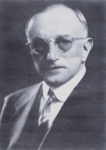
Dr. Charles Elsberg
But step into an operating room at the Neurological Institute a hundred years ago, and you’d meet its first director, Dr. Charles Elsberg.
An expert surgeon, Dr. Elsberg pioneered a way to safely remove tumors from inside the spinal cord.
His innovation was to have the patient’s own body handle the portion of the surgery that would otherwise be too dangerous.
Here’s how it worked: Dr. Elsberg performed the operation in two stages. In the first stage, he made a small opening right over the tumor in the tight membrane that covers the spinal cord. Then he closed the patient’s incision, and the two of them waited. For a week.
During that week, deep inside the body, the incision in the membrane over the spinal cord remained open. This membrane normally fits snugly over the spinal cord. A tumor that grows inside the spinal cord can take up room when there isn’t much to spare, producing a lot of extra pressure inside the membrane.
In Dr. Elsberg’s patients, the body relieved the extra pressure inside the tight membrane by naturally pushing the extra material—the tumor—out through the incision. A week later, when Dr. Elsberg operated again, the tumor would be exposed, free of the spinal cord and ready for him to remove.
Today, Dr. McCormick can safely remove such tumors in a matter of hours, not days. He is heir to the exciting history of intramedullary spinal cord tumor removal here, and he contributes to the field’s continuing development.
Earlier in May, for instance, he spoke to a gathering of spinal neurosurgeons about current intramedullary surgical techniques and their recent technical advances.
As seems fitting for the Director of the Spine Hospital at the Neurological Institute, Dr. McCormick is an internationally recognized expert in intramedullary spinal surgery.
To read about some of Dr. McCormick’s intramedullary spinal cord surgeries, and to watch some videos he produced as teaching tools for other surgeons, take a look at his video page here.
Read more about Dr. McCormick on his bio page here.
Click here to return to The Spine Hospital at The Neurological Institute of New York.
Image Credit: ©Scot Casey/flickr