 Here we bring you another of Dr. Paul C. McCormick’s spinal surgery videos, published by the Journal of Neurological Surgery, Neurosurgical Focus. Each video shows a patient’s spinal problem and the surgery that corrects it. (Read more about this series here.)
The second of Dr. McCormick’s videos shows a rare, but increasingly recognized, spinal problem: spinal cord herniation.
His patient is a woman in her late sixties. For two years she’s been having symptoms of myelopathy, or nerve damage, and her symptoms have been getting worse.
An MRI image reveals a large bend in the woman’s spine. But this obvious bend is not the source of her trouble–it’s just kyphosis, or a rounding of the spine, which isn’t uncommon in older women. No, Dr. McCormick indicates with a tiny blue arrow the miniature bump on the MRI image that spells big trouble for this patient: her spinal cord has herniated out of its protective sheath.
A hernia occurs when part of an organ pouches out through the membranes that normally contain it, into a space it doesn’t belong. The spinal cord (the thin tube of nerves that is protected by the the bones of spinal column) is encased in a very tough membrane called the dura mater. In this patient, a defect in one vertebra has worn a hole in the dura mater, allowing part of the spinal cord to poke out.
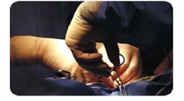
In the video, Dr. McCormick points out the features on the MRI images that led to the correct diagnosis. Then he presents a close-up video of the surgery, explaining what tools he uses, giving tips for handling challenges that might arise during the surgery, and showing how he solves any problems.
For example, as he explains, he needs to separate the spinal cord from the dura mater in the entire area where the cord is bulging out. He starts out on the patient’s right side. But when he moves to the patient’s left, he can’t get at the left side of the problem very well. The spine deviates and turns a little to the right. So, he explains, it will be better to do the whole thing–all the way around–from the patient’s right side.
Dr. McCormick also makes sure to tell his viewers about both the normal and the problematic structures visible at different stages of the surgery. This type of information is extremely helpful for spinal surgeons in training, or for more experienced surgeons who have not dealt with this type of case before. Finally, he describes the patient’s uncomplicated recovery, and shares the postoperative MRIs.
Dr. McCormick notes that he is surprised how often patients request to view a video of the surgery for their condition. And this video is available for anyone to watch–the Journal of Neurological Surgery, Neurosurgery Focus has made it available on YouTube here: Release and repair of a ventral thoracic spinal cord herniation.
But be aware: the video is a graphic and close-up look at surgery. Some patients will want to watch such a detailed video of surgery, but not all will! It should be watched only by interested adults, and with caution.
As spinal cord herniation becomes more widely recognized, many patients and surgeons will likely benefit from this patient’s experience with spinal cord herniation…and from this video made by her experienced surgeon.
Initially posted Nov 19, 2014
Updated Aug 16, 2017