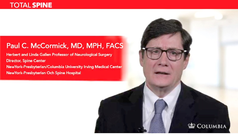
In his latest installment to the Total Spine series of instructional videos, Dr. Paul McCormick describes retropleural thoracotomy, a surgical technique used to treat complex spinal conditions of the thoracic and lumbar spine. “Retropleural thoracotomy is an important...
Astrocytoma
Header | Text |
Summary | An astrocytoma is a tumor that develops from an astrocyte, a type of support cell in the brain and spine. Here at the Spine Hospital at the Neurological Institute of New York, we specialize in spinal astrocytomas. Most spinal astrocytomas are slow-growing and benign–that is, they are not cancer; they will not spread to other parts of the body. However, a small percentage of spinal astrocytomas may grow more aggressively, and a few may even be malignant–cancerous tumors that will spread if left untreated. The type of astrocytoma can give information about the tumor’s possible behavior. For example, pilocytic astrocytomas are tumor types that are usually benign. These astrocytomas are more common in children than in adults. Intermediate tumors like diffuse astrocytomas are more difficult to classify with regard to “benign” and “malignant,” but they are generally slow-growing, though over time they may grow more quickly and become more aggressive. Anaplastic astrocytomas grow at an intermediate rate and are considered malignant. Malignant astrocytomas called glioblastomas grow most aggressively. As spinal astrocytomas grow, they often weave in and out of the tissue of the spinal cord around them. This is called “infiltrating” the spinal cord. Because many astrocytomas infiltrate the healthy spinal cord, it may not be possible to remove them completely without causing damage to the spinal cord. The surgeon’s goal is to remove as much tumor as possible while preserving the maximum of spinal function. The extent of the surgery must be determined during the surgery. The prognosis for an astrocytoma depends on the tumor’s aggressiveness, the type of astrocytoma, and the tumor’s size, location and spread. However, most astrocytomas are very benign and slow growing and can be managed over time with minimal effect on spinal cord function. Similar tumors known as ependymomas arise from other support cells, called ependymal cells, in the brain and spinal cord. Unlike astrocytomas, ependymomas tend not to infiltrate the surrounding spinal cord tissue. In patients older than 20, ependymomas are the more common tumor type, while in patients under 20, astrocytomas are the more common type. Both tumors can occur at any spinal level, though astrocytomas of the lower lumbar and sacral spinal canal are extremely rare. Symptoms and imaging tests cannot always distinguish between astrocytomas and ependymomas. Therefore, it is not uncommon to need biopsy results to make a definitive diagnosis. |
Symptoms | Exact symptoms depend on the location of the tumor and its size. Pain at the site of the tumor is a common early symptom. Weakness, clumsiness, and pins-and-needles in the arms and legs, trouble walking, and incontinence can also develop as the spinal astrocytoma grows. Symptoms usually develop slowly, but in rare cases they may progress quickly. |
Causes and Risk Factors | The causes of astrocytoma are still being researched. Astrocytomas can develop in people of all races, ages, and sexes. They are slightly more common in men and in people with Caucasian ancestry. |
Tests and Diagnosis | Patients who have symptoms suspicious for an astrocytoma will receive a physical examination and answer questions about their symptoms. Doctors may wish to test patients’ nerve functions. They may use imaging scans to help diagnose the tumor and pinpoint its location, size, and spread. Imaging tests helpful for diagnosing astrocytoma include magnetic resonance imaging (MRI) or computed tomography (CT–a CAT scan). A surgical biopsy may also be performed to help determine the type of tumor. |
Treatments | Treatment for astrocytoma consists of microsurgery (open surgery using a surgical microscope and very fine instruments) to remove as much tumor as possible. Sophisticated neurophysiological monitoring may assist the surgeon and help make the surgery safer and more effective. However, astrocytomas may not be distinct from the surrounding spinal cord, so it may not be possible to remove them completely. Therefore, astrocytoma removal surgery is individualized. The neurosurgeons at the Spine Hospital at the Neurological Institute of New York use their extensive training and experience to determine how much tumor may be removed while safeguarding the spinal cord. Postoperative radiation therapy may be recommended for some patients–for example, for patients with aggressive tumors that cannot be fully removed. In most patients, long-term tumor control can be achieved and spinal cord function can be preserved. See the video below of Dr. McCormick performing the procedure.
|
Preparing for Your Appointment | Dr. Paul C. McCormick is an expert in treating astrocytomas. He can also offer you a second opinion. |
Helpful Surgery Overviews
Dr. McCormick will choose the treatment method specific to each patient and situation. Some of the condition’s treatment options may be listed below.
Recent News
Columbia Neurosurgeons Named New York’s Super Doctor
https://www.neurosurgery.columbia.edu/news/columbia-neurosurgeons-named-new-yorks-super-doctor
Dr McCormick video on microsurgical resection of a synovial cyst causing severe lumbar spinal stenosis published in Operative Neurosurgery
Synovial Cysts are a benign and fairly common condition in adult patients. They can arise in most joints throughout the body. In most cases they are asymptomatic but in the spine they can enlarge to produce severe spinal stenosis and pressure on the spinal nerves...
Dr. McCormick invited speaker at 37th Annual Meeting of the Joint Section on Disorders of the Spine and Peripheral Nerves
Dr. McCormick was a featured speaker at the recent 37th Annual Joint Spine Section Annual Spine Summit meeting held in San Diego. He presented at the Innovative Technology Special Session: Operative Video Segments. His presentation was entitled “Intramedullary Tumor,...