 The patient is a man in his late 30s. He has been having upper back pain, progressive difficulty walking, numbness in both hands, and worsening fine motor control.
He has come to see Dr. Paul McCormick, director of The Spine Hospital at The Neurological Institute of New York. Dr. McCormick has diagnosed the man with a spinal cord tumor, called an ependymoma. These tumors are usually benign, slow-growing, and completely removable.
However, their location presents a challenge: in order to access and remove this tumor, Dr. McCormick must gently open the spinal cord itself.
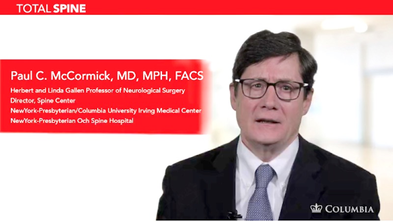
The patient’s surgery is the subject of a video produced by Dr. McCormick and published online recently by the Journal of Neurological Surgery.
In the video, Dr. McCormick reviews the patient’s MRIs which show the tumor itself and two large cysts both above and below it. These cysts, called polar cysts, are hollow. They will collapse and disappear after the tumor is removed. But for now, while the tumor disrupts the normal function of the spinal cord, the cysts are filled with blocked-up spinal fluid.
Dr. McCormick describes the positioning, anesthesia, and spinal cord monitoring he will use during the surgery. The spinal cord monitoring is especially important for an operation like this, in which the surgeon must cut into the spinal cord itself.
“We do have to open the spinal cord to remove these types of tumors,” says Dr. McCormick. “Some numbness in the legs is the most common consequence of this. Removal of these tumors that arise within the actual substance of the spinal cord is challenging. This is because of the delicate nature of the spinal cord and its vital role in every day function, whether it be movement of our arms and legs, the perception of touch, or the identification of pain and temperature stimuli.”
Dr. McCormick narrates the surgery, explaining what clues he uses to find the best spot for opening the spinal cord, what tools he uses to do it, and how he makes sure he is keeping the incision in the exact area he has chosen. He gives similar tips and descriptions of his work at every step of the way: moving the nerves of the spinal cord aside to expose the tumor, isolating the tumor, and separating it from the spinal cord. His expertise and gentle confidence are clear.
The polar cysts above and below this tumor are of great help during this type of operation. Since they are hollow, they give the surgeon some extra room for his instruments. They also let him get a good look at the upper and lower margins of the tumor right away.
“The patient is doing great. He is several years post op,” says Dr. McCormick. “He has some mild numbness in his legs but otherwise functions normally.”
This video is part of a video supplement published online by the Journal of Neurological Surgery: Neurosurgical Focus. In the supplement, Dr. McCormick and other top neurosurgeons share a look through their microscopes with students and doctors both near and far. Dr. McCormick served as editor for the overall 18-chapter supplement. Read more about his work on the project here.
Some patients might enjoy observing the procedure and watching Dr. McCormick’s expert work. Dr. McCormick notes that he is surprised by how often patients request to view a video of the type of surgical procedure they will have. But be aware: this video is a close-up and graphic look at surgery to remove a spinal cord tumor.
If you do want to watch the video and its close-up surgical footage, you can do so here: Microsurgical resection of intramedullary spinal cord ependymoma.


