Dr. McCormick named as one of Castle Connolly’s Top United States and New York Doctors/Neurosurgery for 25 consecutive years.
| Header | Text |
| Summary | Basilar = having to do with the base of the skull Basilar invagination is a condition in which a vertebra (bone) at the top of the spine moves up and back, toward the base of the skull. In this abnormal position, the bone may compress the brain stem and spinal cord. At The Spine Hospital at The Neurological Institute of New York, we specialize in treating basilar invagination. Basilar invagination can be congenital (present at birth) or acquired (develop as a result of injury or disease). To understand more about basilar invagination, it helps to understand a little about the junction between head and neck. This is an area where many important structures come together: it is also the junction between the nervous system tissue of brain and spinal cord, as well as between the bones of the skull and spine. The brain is protected by the bones of the skull, while the spine is protected by the bones of the spinal column. The brain stem is a part of the brain that emerges from its base. It is long and roughly cylindrical–like a stem–and it provides the connection between brain and spinal cord. It leaves the skull through a circular opening in the skull base called the foramen magnum. The brain stem then connects with the spinal cord, which travels down the spinal column in the neck (and beyond). The spine in the neck is called the cervical spine. The individual bones of the cervical spine, called vertebrae, are named according to a simple pattern. The names all start with the letter “C” for cervical, followed by a number that indicates their position. C1 is at the very top of the spine, just below the skull. C1 supports the weight of the skull. C2, the second vertebra, is underneath C1. The joint between C1 and C2 is unusual–it allows much more movement than any other spinal joint. This is the joint that allows the head to turn, rotate and nod. One special feature of this joint is a peg of bone, about the size of the tip of a pinky finger, that sticks up from the front of C2 and fits into a groove in C1. It’s called the dens, or odontoid process. In basilar invagination, C2 and the dens move out of alignment: back and up, toward the foramen magnum. In this position, the dens may compress the brainstem and/or the top of the spinal cord. Like Chiari malformation, a similar condition, basilar invagination reduces the amount of space available for the brainstem and/or the top of the spinal cord. |
| Symptoms | Symptoms vary greatly. The type of symptoms and their severity depend on whether and to what extent the spinal cord, brain stem, spinal nerves, and blood supply are affected. Some cases of basilar invagination cause no severe symptoms. At the other extreme, basilar invagination that puts pressure on the lower brainstem can be fatal. Symptoms may include headache, dizziness, confusion, trouble swallowing, weakness, numbness, and electric-like tingling when the neck is bent. Symptoms usually become worse when the neck is bent, as this position pulls the spinal cord over the projection of the dens. |
| Causes and Risk Factors | Basilar invagination can be either congenital (present at birth) or acquired (developing during a person’s life). Congenital basilar invagination is found more frequently in people with Chiari malformation. Acquired basilar invagination can be the result of rheumatoid arthritis or other diseases. Injury can also lead to basilar invagination. Typical injuries that lead to this condition include bicycle accidents, diving accidents, and falls. |
| Tests and Diagnosis | To diagnose basilar invagination, a physician will order some type of imaging study of the neck and skull base. These may include:
On these images, physicians may draw certain imaginary lines that can help determine how far out of position the dens is. These lines include Chamberlain’s line (between the roof of the mouth and the foramen magnum), McGregor’s line (between the roof of the mouth and the curve at the back of the skull) and McRae’s line (across the opening of the foramen magnum). A dens that projects certain distances over these lines is diagnostic for basilar invagination. |
| Treatments | Every case of basilar invagination is different. Some patients without symptoms may be treated without surgery, using some combination of physical therapy, nonsteroidal anti-inflammatory medications (NSAIDs), and/or a neck brace called a cervical collar. For basilar invagination that causes symptoms, the treatment is usually surgery. The goals of surgery are to
Decompression may be performed using an anterior approach (from the front) or a posterior approach (from behind). Decompression procedures include the anterior odontoidectomy, the removal of the C2 peg. Stabilization is performed with a posterior approach. In the stabilization procedure, called an instrumented spinal fusion, hardware like screws and rods are placed in the joint to fix it, or render it immovable. Transplanted pieces of bone called bone grafts are also placed in the area. The grafts are encouraged to fuse, or grow together, with bone native to the area. This type of procedure can result in a very stable joint. |
| Preparing for Your Appointment | Drs. Paul C. McCormick, Michael G. Kaiser, Patrick C. Reid and Richard C.E. Anderson (Pediatric) are experts in treating basilar invagination. Each can also offer you a second opinion. |
Helpful Surgery Overviews
Dr. McCormick will choose the treatment method specific to each patient and situation. Some of the condition’s treatment options may be listed below.
Recent News
Dr. McCormick named as one of Castle Connolly’s Top United States and New York Doctors/Neurosurgery for 25 consecutive years.
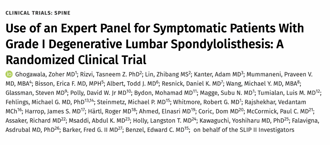
Dr. McCormick participated as both a member of the expert panel as well as a co-author of recently published randomized study on the treatment of grade 1 degenerative spondylolisthesis. This condition commonly occurs in adult patients and is subject to numerous types...
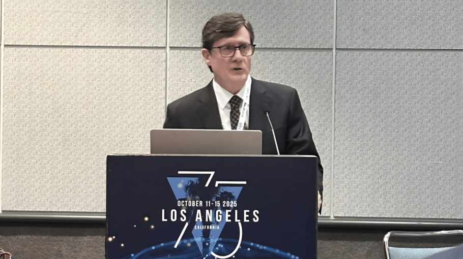
Dr. McCormick was invited to present his experience on the evaluation and microsurgical management of intramedullary spinal cord tumors. Dr. McCormick described his techniques and illustrated on video the safe and complete microsurgical resection of both an...