Chordoma

HeaderText
Summary

A chordoma is a tumor that develops in the bones of the skull or spine. At the Spine Hospital at the Neurological Institute of New York, we specialize in chordomas of the spine.

Spinal chordomas can occur in vertebrae (bones of the spine) at any location along the spinal column. About 1/3 occur at the base of the skull, and about 1/3 occur in the sacrum or coccyx (located at the very base of the spine, between the hips). The remaining 1/3 of chordomas are found at any other spinal level: the cervical spine (neck), thoracic spine (upper and mid-back), or lumbar spine (low back).

These rare tumors are slow-growing. However, they may invade nearby structures, have a tendency to recur after treatment, and may spread to other parts of the body.

Symptoms

As they grow, chordomas may cause bone pain that is not relieved by rest. When the tumors compress nearby nerves, depending on their location, they may cause pain, numbness, or weakness in the arms or legs; changes in bowel and/or bladder function; headache; or double vision.

Causes and Risk Factors

Chordomas develop from remnants of the notochord, a structure that appears early in embryonic development and guides the growth of the bony skull and spine. Normally, some notochord cells form part of the intervertebral discs that act as cushions between the bones of the spine. The rest of the cells are usually replaced by the developing vertebrae. But sometimes, additional notochord cells are enclosed by the developing bones. These notochord cells may one day give rise to a chordoma.
Chordomas can arise in people of any age. Skull base chordomas are more frequent in children and young adults, while spinal chordomas are more common later in life.

A specific change in a gene called brachyury is found in most people with chordomas. The change in this gene cannot be said to cause chordoma by itself — many people in the general population have this specific change, but the vast majority of those people will never develop chordoma. Research is ongoing to identify what might cause chordoma in people who are predisposed by the change in the brachyury gene.

The brachyury gene is found in mammals, fish, and many other organisms, and plays an important role in developing an organism’s midline. The midline, or center line of symmetry, includes the spine in humans; in animals with tails, the midline includes the tail. Brachyury was first identified as a gene that affects tail length in mice, and this discovery is reflected in its name: from the Greek, brakhus = short; oura = tail.

Tests and Diagnosis

As with all spine tumors, imaging studies are essential in diagnosing chordomas. Because these tumors often involve bony structures, computed tomography (CT) scans are used to assess the degree of bony involvement. Magnetic resonance (MR) scans also are used to distinguish the tumor from the sensitive structures around it. For both CTs and MRs, an agent like a dye may be administered intravenously so that neurosurgeons can better visualize the tumor against the normal tissue in the background.

Spinal angiography, which reveals arteries and vascular structures around the tumor, may also be used. This imaging study can help direct the surgical approach to the tumor by showing which blood vessels it affects.

Treatments

The goal of chordoma surgery is complete tumor removal. However, the location of these tumors and their involvement with important structures in the body may make complete removal difficult or impossible.

The neurosurgeons at the Spine Hospital at the Neurological Institute of New York tailor a surgical plan to each individual patient. Complete removal, when possible, may involve combined or multiple surgical procedures or approaches. Spinal fusion and stabilization is often performed following tumor removal. Possible surgical procedures include:

Stereotactic radiosurgery, which uses a highly focused beam of radiation to target the cancer cells, may be used along with traditional open surgery. However, chordomas are notoriously resistant to chemotherapy and standard radiation therapy. Long term tumor control or cure can often be attained with complete tumor resection and adjuvant (post-surgical) radiosurgery in select cases.


Preparing for Your Appointment

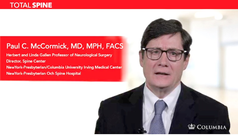
Dr. Paul C. McCormick is an expert in treating chordoma. He can also offer you a second opinion.

Helpful Surgery Overviews

Dr. McCormick will choose the treatment method specific to each patient and situation. Some of the condition’s treatment options may be listed below.

Recent News