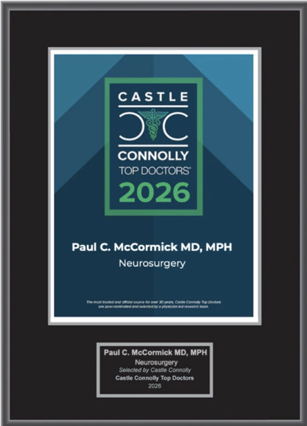
Dr. McCormick named as one of Castle Connolly’s Top United States and New York Doctors/Neurosurgery for 25 consecutive years.
| Header | Text |
| Summary | Spinal= having to do with the spine or spinal cord The meninges are the protective membranes that enclose the brain and spinal cord. At The Spine Hospital at The Neurological Institute of New York, we specialize in spinal meningiomas, or tumors that arise in the meninges around the spinal cord. Meningiomas are usually benign (noncancerous), slow-growing tumors, although in rare cases they may be malignant and invade surrounding tissue. Meningiomas account for approximately 25% of all tumors that involve the spinal canal. They can occur at any location throughout the spine, but are predominantly found in the thoracic (mid-spine) region, probably because this is the largest segment of the spine. |
| Symptoms | The symptoms of a spinal meningioma are caused by the pressure that the growing tumor exerts on the spinal cord or spinal nerves. Depending on the location of the tumor, meningiomas may cause weakness or numbness in the arms or legs, and/or difficulty with bladder, bowel and/or sexual function. The symptoms tend to become more severe as the tumor grows in size. |
| Causes and Risk Factors | The exact causes of spinal meningiomas are not well understood. Spinal meningiomas occur approximately four times as often in women as in men. However, the rare malignant form of meningioma is more common in men than women. The average age at diagnosis is 45 years. People with the inherited disorder neurofibromatosis II (NF 2) are more prone to developing meningiomas. |
| Tests and Diagnosis | Imaging studies are the key component in the diagnosis of meningiomas. Magnetic resonance (MR) scans provide useful detail about the tumor’s size, location, and effect on surrounding structures. MR scans use magnets, radio waves, and computer technology to produce images of organs and tissues like the brain and spinal cord. The scans may also be taken after injecting a contrast dye that highlights the tumor tissue against the background of normal tissue. Computed tomography (CT) can be useful in patients with pacemakers or other metallic devices, who cannot undergo MR imaging. CT uses a combination of X-rays and computer technology to produce detailed images of bones and soft tissues. Calcium deposits, common in meningiomas, are more precisely identified by CT scan. |
| Treatments | If a meningioma is small and does not cause symptoms, it may be observed over time rather than removed. In cases where treatment is necessary, the first line of treatment is usually surgery. Because the tumors are typically benign–that is, they are not cancerous and do not spread–complete removal often results in a cure. Some tumors, however, may be difficult to remove completely. These include tumors with unusual amounts of calcium, tumors that are located in difficult areas to access, or tumors that invade surrounding tissue. Some meningiomas may recur, even in cases of complete removal. Stereotactic radiosurgery, a non-surgical form of radiation therapy, may be used as a follow-up treatment for tumors that cannot be totally removed or for those that re-grow. To see a video of Dr. McCormick performing Spinal Meningioma surgery, click the video link below. |
| Preparing for Your Appointment | Drs. Paul C. McCormick, Michael G. Kaiser, Alfred T. Ogden, Christopher E. Mandigo, Patrick C. Reid, Richard C.E. Anderson (Pediatric), and Neil A. Feldstein (Pediatric) are experts in treating spinal meningiomas. They can also offer you a second opinion. |
Helpful Surgery Overviews
Dr. McCormick will choose the treatment method specific to each patient and situation. Some of the condition’s treatment options may be listed below.
Recent News
Dr. McCormick named as one of Castle Connolly’s Top United States and New York Doctors/Neurosurgery for 25 consecutive years.

Dr. McCormick participated as both a member of the expert panel as well as a co-author of recently published randomized study on the treatment of grade 1 degenerative spondylolisthesis. This condition commonly occurs in adult patients and is subject to numerous types...
Dr. McCormick was invited to present his experience on the evaluation and microsurgical management of intramedullary spinal cord tumors. Dr. McCormick described his techniques and illustrated on video the safe and complete microsurgical resection of both an...