| Header | Text |
| What is a Pedicle Subtraction Osteotomy (PSO)? | Pedicle = a section of bone that connects the front and back of a vertebra
Subtraction = removal
Osteotomy = procedure in which bone is cut
A pedicle subtraction osteotomy (PSO) is a surgical procedure used in adults and children to correct certain deformities of the spine. A spine with too much kyphosis (forward curvature, or “hunchback”) or too little lordosis (inward curvature, or “swayback”) can be corrected with a PSO.
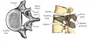
During a pedicle subtraction osteotomy, the shaded areas of bone are removed. The spine is then realigned and stabilized in its new alignment.
For general information about spinal deformity and its correction, please see the deformity correction overview page.
|
| When is this Procedure Performed? | When viewed from the side, a typical spine has a few gentle curves that balance each other out, aligning the body’s center of gravity over the pelvis. This alignment is called sagittal balance. Conditions like hyperkyphosis, ankylosing spondylitis, and flatback syndrome can put the body’s center of gravity too far ahead of the pelvis, resulting in sagittal imbalance. Pedicle subtraction osteotomy is one surgical option for treating sagittal imbalance.
Viewed from behind, a typical spine is straight. Side-to-side curvature visible from behind is known as scoliosis. A moderate amount of scoliosis can sometimes be corrected during a pedicle subtraction osteotomy.
Spinal curvature is measured in degrees. For example, a side-to-side curvature of 10 degrees or more qualifies as scoliosis. The curve of kyphosis in an average spine measures between 20 and 40 degrees; a kyphosis of 70 degrees or more (technically called a hyperkyphosis) may be treated with surgery.
A PSO yields about 30 degrees of correction. It is well-suited for treating sharp, angular kyphosis, some cases of flatback syndrome, and some cases of ankylosing spondylitis. It is most often performed in the lumbar (lower) spine. It is sometimes coupled with one or more osteotomies to achieve an even greater degree of correction.
Compared to the other deformity correction procedures, a PSO removes a moderate amount of bone and allows a moderate amount of realignment. It removes the facet joints at the back of the target vertebra and the vertebrae above and below, the pedicles that attach the back of the target vertebra to its front, the laminae, and also a portion of the target vertebral body.

Image credit:
© [Henry Vandyke Carter (1831-1897)] / Public Domain
|
| How is this Procedure Performed? | A pedicle subtraction osteotomy is performed under general anesthesia, which means the patient is unconscious. Spinal cord monitoring techniques like SSEP (somato-sensory evoked potentials) and MEP (motor-evoked potentials) measure electrical activity in nerves and help ensure the safety of the spinal cord throughout the operation.
Once the patient is unconscious, he or she is carefully placed face-down on a special hinged operating table. The table is set up something like an upside-down flattened letter “V”, where the tip of the V points up towards the ceiling. (The table is much flatter than an actual upside-down letter “V”.) An incision is made in the center of the back and the bones of the spinal column are exposed.
Then the surgeon places screws in the vertebrae above and below the targeted area. These screws are called pedicle screws because they are inserted into the pedicles–the thick, sturdy columns of bone that connect the back of the vertebra with the front. Later in the surgery, the pedicle screws above and below the target vertebra will provide attachment points for the rods that will hold the spine in position while it heals.
Next the surgeon removes the projections, called processes, from the back of the target vertebra. The surgeon hollows out a space under the pedicles of the target vertebra, removing the pedicles. Then the surgeon enlarges the space, creating a wedge-shaped hollow space in the vertebra.
Once the wedge-shaped hollow is complete, its upper and lower portion can be brought together. This closes the wedge and provides about 30 degrees of correction to the deformity. The surgeon gradually widens the special surgical table from its reverse “V” into a flatter position, until the bone surfaces lie against each other and the wedge is closed.
Finally, rods are placed in the pedicle screws to help hold the spine in position while it heals. As the bone surfaces of the upper and lower wedge grow together, permanently fusing into one solid bone, they will provide lasting strength and stability to the vertebra.
|
| How Should I Prepare for this Procedure? | Before any deformity correction surgery, it is important to stop using tobacco products. Nicotine has a very detrimental effect on bone fusion and surgery outcomes. If you currently smoke or use tobacco, speak to your neurosurgeon about quitting.
Be sure you understand the goals of your or your child’s surgery, the risks, and what can be expected from the recovery period. If you have any questions at all about the procedure or the recovery, speak with your or your child’s neurosurgeon. To make it easier, write down your or your child’s questions as they arise, and bring the list to your appointments.
Make sure to tell your doctor about any medications or supplements that you or the patient are taking, especially medications that can thin your blood such as aspirin. Your doctor may recommend you or the patient stop taking these medications before your procedure. To make it easier, write all medications down before the day of surgery.
Be sure to tell your doctor if you or the patient have an allergy to any medications, food, or latex (some surgical gloves are made of latex).
On the day of surgery, remove any nail polish or acrylic nails, do not wear makeup and remove all jewelry. If staying overnight, bring items that may be needed, such as a toothbrush, toothpaste, and dentures. You will be given an ID bracelet. It will include your name, birthdate, and surgeon’s name.
|
| What Should I Expect After the Procedure? | How long will I stay in the hospital?
Most patients stay in the hospital approximately 3-7 days.
Will I need to take any special medications?
Postoperative discomfort will be controlled with pain medication.
Will I need to wear a brace or collar?
If the pedicle subtraction osteotomy is in the neck, patients are often prescribed a cervical (neck) collar. Otherwise, no brace is usually necessary.
When can I resume exercise?
Low impact exercise can typically resume after 3 months.
Will I need rehabilitation or physical therapy?
Physical therapy can be beneficial for gait training / ambulation (walking).
Will I have any long-term limitations due to a pedicle subtraction osteotomy?
Some patients will notice decreased mobility due to fusion. Speak with your surgeon about what you can expect after your particular surgery.
|
| Preparing for Your Appointment | At The Spine Hospital at the Neurological Institute of New York, Drs. Paul C. McCormick, Michael G. Kaiser, Peter D. Angevine, Christopher E. Mandigo and Patrick C. Reid are experts in pedicle subtraction osteotomy.
Dr. Richard C. E. Anderson is an expert in pedicle subtraction osteotomy for pediatric patients.
|


